


Dermatophytosis is a fungal infection affecting the superficial layer of the epidermis and of skin keratinized adnexa (i.e. nails and hairs), which is caused by a group of fungi referred to as “dermatophytes”. The fungal group responsible for dermatophytosis include three genera: Microsporum, Epidermophyton and Trichophyton.
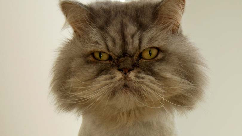
Dermatophytosis in cats is usually caused by three species of dermatophytes: Microsporum canis (98% of cases), Microsporum gypseum and Trichophyton mentagrophytes. The importance of dermatophytosis in cats (particularly as regards those forms caused by species of Microsporum) is related not only to its high incidence in cats and the high frequency with which it is misdiagnosed, but also to the risk of potential transmission to human.
There are several factors which can predispose cats to the infection by dermatophytes. These can be categorized in (1) environmental factors, such as hot-humid environments, and (2) subjective factors such as young age, genetic predisposition, any condition which can reduce the body’s defense mechanisms (for example, malnutrition, parasitic diseases, chronically debilitating diseases and virus-caused immunodeficiency states) and any condition affecting the integrity of the skin. Predisposed subjects also include cats with long hair (perhaps because of the difficulty to remove fungal spores by self-licking the hair), as well as cats who are often bathed (with removal of the sebum which have antifungal properties).
In order to understand the possible origin of an infection, it is important to remember that, as a general rule, Microsporum canis is a “zoophilic” fungus (literally, an animal-loving fungus) while Microsporum gypseum is a “geophilic” fungus (literally, a soil-loving fungus). Therefore the infection by M. canis will be transmitted more frequently through direct contact with infected or carrier animals, while the infection by M. gypseum will occur more commonly following contact with a contaminated environment (particularly the contaminated soil). Regardless of the modality of infection, after the fungal spores adhere to the skin or the hair, they can germinate and invade the keratinized layers of the skin, nail and hair. The penetration into the keratinized tissues is supported by the production of keratinolytic enzymes by the fungal hyphae (growing vegetative fungal stages). The penetration continues until the fungus reaches the keratin-producing layer of the epidermis, at which point a sort of balance between fungal growth and keratin production develops. This infection is usually self-limiting, since fungal forms are automatically eliminated from the body when the infected hairs fall out or the infected skin exfoliates. In addition, the infection can be also limited or even eliminated when the inflammatory reaction of the host is particularly effective.
Dermatophytosis in cats can have very different clinical pictures, depending on the interaction of the host with the fungus. A common symptom is the weakening or loss of the hair, accompanied by the appearance of skin areas with fragile or broken hairs or even of areas of alopecia, with or without skin erythema. Skin exfoliation is generally marked, characterized by the presence of “ash-like” scales, and it is sometimes accompanied by the formation of crusty material. Commonly affected areas include the muzzle, the head and the limbs. When the inflammatory reaction is particularly intense, pimples and pustules can be present. Pruritus is variable, while secondary bacterial infections are not frequent in cats. A particular clinical picture of dermatophytosis in cats is the so-called pseudomycetoma, which appears as a non-painful, hard lump affecting the deeper layers of the skin (dermis and ipodermis) along the torso or at the base of the tail. This kind of lesion develops in cats which are not protected by an efficient immune system, for example in cats affected by FIV or FeLV.
Once properly diagnosed, the therapeutic protocol for dermatophytosis in cats must include the use of systemic drugs (e.g., griseofulvin, terbinafine, itraconazole) in combination with topical drugs (e.g., clotrimazole, sulconazole, chlorexidine). The systemic treatment, administered in oral formulation, should continue for two weeks after the disappearance of clinical signs and until the results of cultural tests are negative. As a general rule, when clinical lesions persist after eight weeks of treatment, the presence of a drug-resistant fungus or of an underlying primary disorder must be suspected. The topical treatment, which should be always associated with a systemic treatment to be effective, can be administered in various formulations such as solutions, gels, ointments and creams.
Other possible measures include clipping, at least around the area that is affected, as well as certain appropriately prepared vaccines which can reduce and significantly prevent the appearance of symptoms although not providing full protection from infection.
The therapeutic protocol for dermatophytosis in cats should also include an environmental decontamination program with appropriate disinfectants. In fact it should not be forgotten that the environment can be always an infection or re-infection source for cats, as well as for humans.
Unfortunately dermatophytosis in cats is too often misdiagnosed, poorly treated and underestimated in its potential zoonotic risk (risk of transmission to humans); therefore it is the responsibility of the veterinarian not to diminish the importance of this risk and to define effective prophylactic and/or therapeutic plans to properly treat and control the disease, while it is the responsibility of the cat owner to follow these plans and strictly adhere to the instructions given by the veterinarian.